Cervical Spine (Neck) CT Scan
Current as of: December 9, 2019
Author: Healthwise Staff
Medical Review: Adam Husney, MD - Family Medicine & Martin J. Gabica, MD - Family Medicine & Howard B. Schaff, MD - Diagnostic Radiology
Cervical Spine (Neck) CT ScanCourtesy of Intermountain Medical Imaging, Boise, Idaho. All rights reserved. 
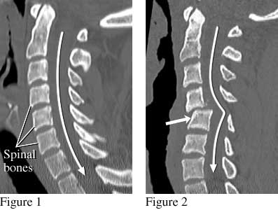
The side view of the spine (figure 1) shows the normal position of the spinal bones. The spinal cord (not visible in this image) follows the path of the long white arrow. Figure 2 shows shifted spinal bones (short thick arrow) from a fracture that damaged the spinal cord. This person was paralyzed after being ejected during an automobile crash while not wearing a seat belt. Current as of: December 9, 2019 Author: Healthwise Staff Medical Review: Adam Husney, MD - Family Medicine & Martin J. Gabica, MD - Family Medicine & Howard B. Schaff, MD - Diagnostic Radiology |
Search the
Healthwise Knowledgebase This information does not replace the advice of a doctor. Healthwise, Incorporated, disclaims any warranty or liability for your use of this information. Your use of this information means that you agree to the Terms of Use. Learn how we develop our content. To learn more about Healthwise, visit Healthwise.org. © 1995-2020 Healthwise, Incorporated. Healthwise, Healthwise for every health decision, and the Healthwise logo are trademarks of Healthwise, Incorporated. |